Smiling woman with a blonde bob and tan cardigan showing healthy teeth after periodontal treatment

CBCT

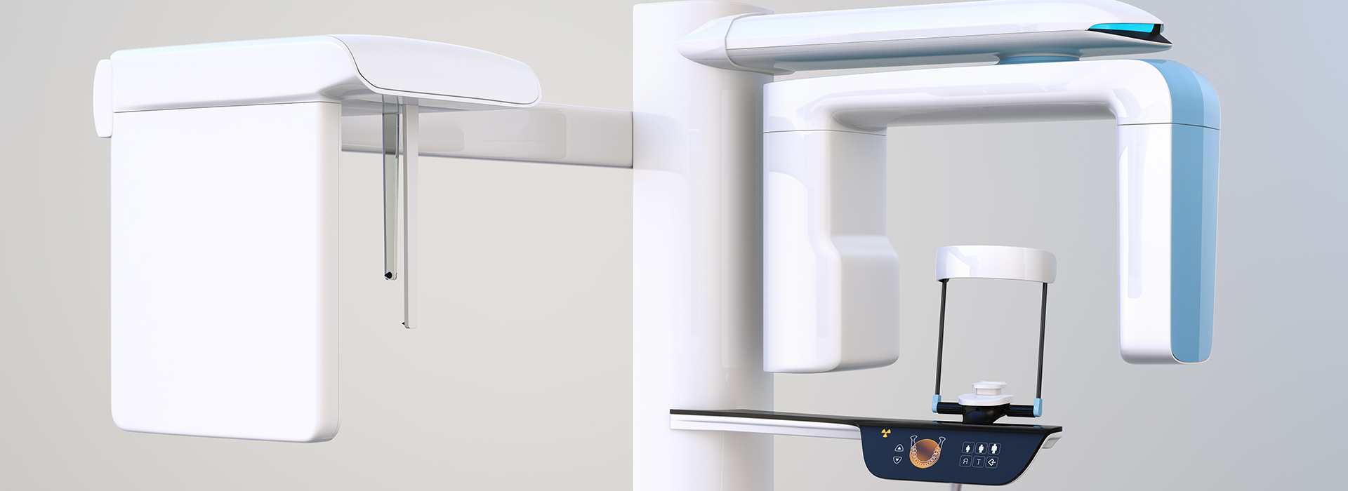
White and blue CBCT dental scanner with a chin rest and control panel used for 3D dental imaging

At the office of Macri Dental, we use the latest technology to provide our patients with the most precise and gentle care. We firmly believe that achieving successful treatment outcomes relies on using the most advanced diagnostic technology. By obtaining high-resolution and detailed 3D images, we can develop the right treatment plan and provide the highest level of care.

As a state-of-the-art practice, we utilize one of the most advanced CBCT (cone-beam computed tomography) imaging systems to obtain the sharpest 3D diagnostic films while exposing the patient to the lowest level of radiation.

CBCT provides leading-edge diagnostics and facilitates precision care

With the advanced and versatile capabilities provided by our CBCT imaging system, it’s quick, safe, and comfortable to obtain detailed 3D views of a patient's teeth, jaws, and surrounding anatomy.

Multiple applications to support optimal treatment outcomes

Because they obtain targeted, detailed, and distortion-free views from multiple angles, CBCT scans provide essential information for numerous analyses and the assessment of maxillofacial disorders or pathology. With applications in every area of dental care, CBCT imaging facilitates precise surgical planning, including the accurate placement of dental implants.

Experienced and trusted care for healthy and beautiful smiles

We value your time as well as your investment in oral health. Our office maintains an unwavering commitment to excellence and strives to provide the most advanced and comfortable solutions to meet your family’s dental needs. With patient care and comfort as our top priorities, you can depend on our practice for the highest quality of skilled and compassionate care.


Frequently Asked Questions

What is cone-beam computed tomography and how does it differ from traditional dental X-rays?

+

Cone-beam computed tomography, commonly called CBCT, is a three-dimensional imaging method that captures a volumetric dataset of the teeth, jawbone, and surrounding structures. Unlike traditional intraoral X-rays or panoramic films that produce flat, two-dimensional images, CBCT allows clinicians to view anatomy in axial, coronal, and sagittal planes and to render a manipulable 3D model. This additional dimensionality reveals details such as root morphology, bone contours, and spatial relationships that can be hidden on 2D films.

Because CBCT provides a true volume rather than a compressed projection, clinicians can rotate, slice, and zoom into regions of interest to better characterize complex anatomy. That capability reduces estimation and guesswork during diagnosis and planning, which often leads to more precise and predictable treatment decisions. The technology complements rather than replaces conventional films, with each modality used according to the diagnostic need.

Why might my dentist recommend a CBCT scan for my care?

+

A dentist may recommend CBCT when two-dimensional imaging is insufficient to answer specific clinical questions or when precise spatial information is required for safe treatment. Common reasons include implant planning, assessment of complex root canal anatomy, evaluation of pathology, pre-surgical assessments, and detailed examination of the temporomandibular joints and airway. In these situations, CBCT can clarify anatomy that would otherwise remain uncertain on 2D images.

By providing objective measurements of bone volume, nerve position, and the relationship of roots to anatomical landmarks, CBCT helps clinicians select the safest and most appropriate techniques. It also aids in communicating findings and treatment options to patients and collaborating specialists, promoting informed decision-making. Ultimately, the scan is used only when it adds clear diagnostic value to the care plan.

Is CBCT safe and how is radiation exposure managed?

+

Dental CBCT units are specifically engineered for head and neck imaging and, when used with appropriate field-of-view and exposure settings, typically deliver lower radiation doses than conventional medical CT scans. Practices follow the ALARA principle—"as low as reasonably achievable"—meaning each scan is justified based on clinical need and optimized to minimize exposure. Modern machines also allow clinicians to limit the scanned volume to the region of interest, which reduces dose compared with full-volume scans.

Before any scan, the dental team reviews your health history and prior imaging to confirm that CBCT is the most appropriate test. Technicians position and instruct patients to reduce motion, and operators choose exposure parameters that balance image quality with dose reduction. If you have specific concerns about radiation, discuss them with your provider so they can explain the rationale and safety measures in your case.

What should I expect during a CBCT appointment?

+

A CBCT appointment is generally quick and comfortable. You will be asked to sit or stand and to remain still while the imaging arm rotates around your head, with most dental scans completed in less than a minute of exposure time. Staff members will position you carefully, provide instructions to minimize movement, and remain nearby to offer reassurance throughout the brief exam.

There is no special preparation for most patients beyond removing metal jewelry or removable dental appliances that could create artifacts. The experience is similar to routine radiographs in terms of comfort, and staff will answer any questions before starting. If you have mobility concerns, claustrophobia, or other special needs, let the office know in advance so accommodations can be made.

How is CBCT used for implant planning and oral surgery?

+

CBCT is a cornerstone of modern implant planning because it provides accurate measurements of bone height, width, and density while showing the position of critical anatomical structures such as the inferior alveolar nerve and maxillary sinuses. These details allow clinicians to determine whether a site has sufficient bone for an implant, choose the ideal implant size and angulation, and identify areas that may need grafting or modification. CBCT datasets can also be used to design and fabricate surgical guides that translate the digital plan into precise intraoperative placement.

Using 3D imaging reduces intraoperative surprises and supports safer, more predictable outcomes by informing decisions before the first incision. It also facilitates communication with surgical teams and laboratories when multidisciplinary care is required. At Macri Dental we integrate CBCT data into our digital workflows to enhance planning accuracy and patient education.

How can CBCT imaging help with root canal diagnosis and retreatment?

+

CBCT often reveals root canal anatomy and pathology that are not visible on conventional periapical films, such as additional canals, unusual canal curvature, fractures, or the full extent of periapical lesions. By examining the tooth and surrounding structures in three dimensions, clinicians can identify treatment obstacles and better plan endodontic procedures or revision treatments. This detailed visualization can be critical when symptoms or previous treatment outcomes are unclear.

When retreatment is considered, CBCT can show the presence and location of missed canals, perforations, and the relationship of lesions to adjacent teeth or anatomical spaces. That information helps determine whether nonsurgical retreatment, surgical endodontic intervention, or referral to a specialist is the most appropriate option. Interpretation is combined with clinical findings to guide a targeted and conservative approach.

What role does CBCT play in evaluating the airway, sinuses, and TMJ?

+

CBCT provides a reliable three-dimensional view of the nasal cavity, paranasal sinuses, airway passages, and temporomandibular joints, allowing clinicians to assess structural contributors to breathing dysfunction, sinus disease, or TMJ disorders. Volumetric airway analysis can identify areas of constriction or anatomical variations that may influence sleep-disordered breathing evaluation, while detailed condylar and joint space images assist in diagnosing degenerative changes or internal derangements. In sinus assessments, CBCT helps locate mucosal thickening, obstruction, and the relationship of dental roots to the sinus floor.

Although CBCT is valuable for anatomical assessment, it is one component of comprehensive care; for example, a sleep study is required to diagnose sleep apnea definitively. Findings from CBCT are integrated with medical history, clinical examination, and other diagnostic tests to form a complete picture. When indicated, results can be shared with medical specialists to coordinate multidisciplinary management.

Who interprets CBCT scans and how are images used to inform treatment plans?

+

CBCT images are interpreted by the treating dentist or a clinician with specialized training in radiographic interpretation, and complex cases may be referred to or reviewed by an oral and maxillofacial radiologist. Proper interpretation requires systematic review of all anatomical regions captured in the volume to avoid overlooking incidental findings that could affect care. Clinicians correlate CBCT findings with clinical exam results and other tests to ensure a comprehensive and accurate diagnosis.

Once interpreted, CBCT data informs decisions about surgical approach, implant positioning, restorative design, or endodontic strategy, and it helps anticipate potential complications. The images also support patient education by allowing clinicians to demonstrate anatomy and explain recommended steps. This evidence-based approach reduces uncertainty and improves the predictability of treatment outcomes.

Can CBCT data be shared with specialists and used in digital workflows?

+

Yes, CBCT datasets are typically saved in DICOM format and can be exported or shared with specialists, laboratories, and digital service providers to support coordinated care. The same volumetric data can be integrated with intraoral scans, CAD/CAM systems, and 3D printing workflows to fabricate surgical guides, prosthetic components, or diagnostic models. This interoperability streamlines communication and ensures that all members of the care team work from the same precise anatomical information.

Sharing CBCT data also facilitates second opinions and collaborative planning for multidisciplinary cases such as orthodontic/implant coordination or complex restorative rehabilitation. Secure transfer methods and clear documentation help protect patient privacy while enabling efficient collaboration. At Macri Dental we use standardized digital exports to coordinate with partners when cases require additional expertise or laboratory fabrication.

How should I prepare for a CBCT scan and are there any limitations I should know about?

+

Preparation for a CBCT scan is minimal: patients are usually asked to remove glasses, jewelry, and removable dental appliances that could create artifacts in the image. It is important to inform the dental team if you are pregnant, have implanted medical devices, or have conditions that could affect positioning or the ability to remain still. Discuss any concerns with staff beforehand so accommodations can be made for comfort and safety.

Limitations of CBCT include susceptibility to motion artifacts, metal scatter from restorations, and resolution constraints for very fine structures compared with some specialized modalities. Small carious lesions or subtle soft-tissue findings may not be as well characterized on CBCT as on other targeted imaging studies. For these reasons, clinicians select CBCT when its benefits outweigh limitations and when the scan clearly advances diagnosis or treatment planning.

Contact Us

Exceptional Dentistry, Centered Around You

Discover a dental experience built around your comfort, needs, and goals. Our team is here to answer your questions, help you explore your treatment options, and make scheduling your visit simple and convenient. Contact our office today and take the next step toward personalized, high-quality dental care for you and your family.

Office Hours

Monday
9:00 am - 5:00 pm
Tuesday
7:00 am - 4:00 pm
Wednesday
7:00 am - 3:00 pm
Thursday
7:00 am - 2:00 pm