
Smiling woman with a blonde bob and tan cardigan showing healthy teeth after periodontal treatment

Digital Radiography

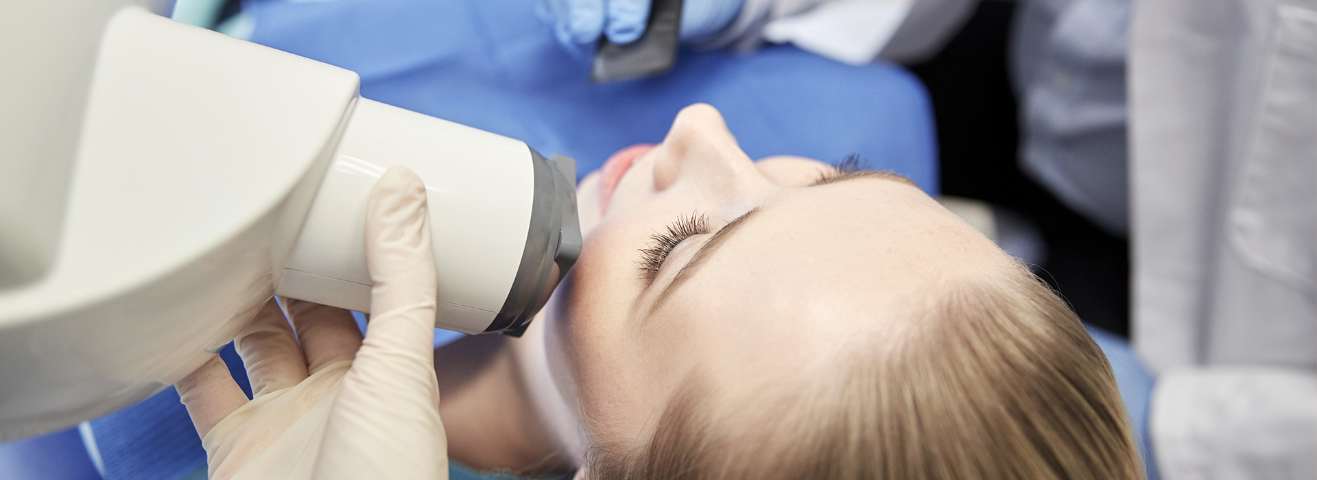
Gloved dental professional positioning a digital X-ray device beside a young patient's face as she reclines in a dental chair

Digital radiography utilizes computer technology and digital sensors for the acquisition, viewing, storage, and sharing of radiographic images. It offers several advantages over the older traditional film based methods of taking x-rays. The most significant of these advantages is that digital radiography reduces a patient’s exposure to radiation. Other benefits are that images can be viewed instantly after being taken, can be seen simultaneously as needed by multiple practitioners, and can be easily shared with other offices. Digital x-rays are also safer for the environment as they do not require any chemicals or paper to develop.

An electronic pad, known as a sensor is used instead of film to acquire a digital image. After the image is taken, it goes directly into the patient’s file on the computer. Once it is stored on the computer, it can be easily viewed on a screen, shared, or printed out.

Frequently Asked Questions

What is digital radiography and how does it differ from traditional X-rays?

+

Digital radiography uses electronic sensors and computer processing to capture dental images instead of photographic film. Images appear on a monitor within seconds of exposure, eliminating darkroom development and allowing immediate review. This rapid availability helps clinicians identify issues quickly and discuss findings with patients during the same visit.

Unlike film, digital files can be enhanced, measured, and compared side-by-side with prior studies to show subtle changes over time. The workflow includes sensor capture, software refinement, and calibrated display, which together produce more consistent image quality. Because the images are digital from the outset, they are easier to store, retrieve, and integrate with other diagnostic data.

How does digital radiography improve diagnostic accuracy?

+

High-resolution digital sensors capture fine anatomical detail that can reveal small cavities, hairline fractures, and early bone changes that might be harder to see on film. Clinicians can adjust contrast, brightness, and magnification to highlight areas of concern and use measurement tools to assess lesion size or bone levels. These tools enhance the clinician's ability to detect and monitor conditions earlier and with greater confidence.

Digital systems also enable consistent, reproducible images that facilitate reliable comparisons from visit to visit. Side-by-side review of current and prior images helps clinicians recognize slow progressions or rapid changes in periodontal disease, restorations, or healing. The improved visual information supports evidence-based decisions and more precise treatment recommendations.

Is digital radiography safer than traditional film X-rays?

+

Digital sensors are more sensitive to X-rays than film, which allows diagnostic-quality images to be obtained with lower radiation exposure. Lower exposure is particularly meaningful for patients who need frequent imaging, children, and those who warrant special precautions. Reduced radiation does not compromise image clarity; modern digital techniques preserve diagnostic detail while improving safety.

Because images can be reviewed immediately, technicians can confirm adequacy on the spot and retake only when necessary, which further limits cumulative exposure. Digital workflows also eliminate chemical processing, reducing handling of potentially hazardous materials and supporting infection-control practices. Together, these factors make digital radiography a safer and more efficient option for routine dental imaging.

What should I expect during a dental visit that uses digital imaging?

+

During a digital imaging appointment, a sensor or film holder is placed briefly in the mouth while a fast exposure is made, similar to traditional X-rays but typically quicker. The resulting image appears on a monitor within seconds, allowing the clinician to review it with the patient and explain any findings in real time. The process is generally comfortable and only adds a few minutes to a standard exam.

If an image needs to be repeated for clarity, the clinician can make small positioning or exposure adjustments immediately, which minimizes repeat exposures. Children and anxious patients benefit from shorter imaging times and the ability to preview images on screen. The practice also uses protective measures such as lead aprons and thyroid collars when indicated to further enhance safety.

How does digital radiography support treatment planning for implants and restorations?

+

Digital radiographs provide detailed views of tooth roots, surrounding bone, and existing restorations that help clinicians assess suitability for implants, crowns, and other restorative work. When combined with three-dimensional imaging and digital impressions, two-dimensional radiographs contribute to comprehensive planning by clarifying bone levels, angulation, and restorative margins. These integrated data allow clinicians to select more conservative, predictable treatment approaches.

Stored digital images also become part of the patient’s longitudinal record, supporting preoperative planning and postoperative monitoring. Precise documentation helps coordinate care with specialists and dental laboratories, ensuring that surgical guides, prosthetics, and restorative components fit as intended. The result is more efficient treatment planning and improved clinical outcomes.

Can digital images be shared with specialists and stored securely?

+

Yes. Digital radiographs can be exported in standard formats and transmitted securely to specialists, labs, or referring clinicians, which speeds consultation and collaborative treatment planning. Electronic transfer preserves image resolution and allows multiple providers to review the same high-quality files without waiting for physical films. Secure sharing reduces administrative delays and supports timely, coordinated care.

The files are stored within encrypted electronic systems that meet contemporary privacy and recordkeeping standards, reducing the risk of lost or damaged films. Authorized members of the care team can retrieve images quickly during appointments, and the digital archive simplifies continuity when patients move or change providers. At Macri Dental, secure storage and controlled access are part of our commitment to patient privacy and clinical efficiency.

Are there special benefits of digital radiography for children and pregnant patients?

+

Because digital sensors require less radiation to produce diagnostic images, they are particularly advantageous for children who may need monitoring over many years. Faster exposures and immediate review reduce the need for repeat imaging, which also helps limit cumulative dose. Pediatric imaging workflows can be adapted to make the process quick and comfortable for younger patients.

For pregnant patients, dental X-rays are generally avoided unless absolutely necessary, but when imaging is required digital systems help minimize exposure and allow the team to take appropriate protective measures. Clinicians weigh the diagnostic benefits against potential risks and use shielding and positioning strategies to protect the abdomen and thyroid. Clear communication about necessity and safety helps patients feel informed and reassured.

How does the practice ensure image quality and consistency over time?

+

Quality starts with properly calibrated displays, regularly maintained sensors, and standardized exposure protocols that the clinical team follows for each image type. Routine equipment checks and software updates help preserve image fidelity and ensure measurements remain accurate across visits. Staff training on positioning and technique further reduces variability and enhances reproducibility.

Consistent image capture and robust storage practices also make longitudinal comparisons meaningful, allowing clinicians to detect slow trends or sudden changes with confidence. When needed, images can be annotated and archived within the patient record to document findings and treatment decisions. The office of Macri Dental emphasizes these protocols to support reliable diagnostics and continuity of care.

Do digital radiographs reduce environmental impact compared with film?

+

Yes. Digital radiography eliminates the need for film, chemical developers, and disposable packaging associated with traditional radiographic processing. Removing chemical processing reduces hazardous waste and the environmental burden of disposal, and it decreases the practice’s dependence on single-use materials. Many practices adopt digital imaging as part of broader sustainability efforts.

Beyond waste reduction, digital systems can lower the carbon footprint tied to shipping and storing physical films by enabling electronic exchange and centralized archives. Fewer repeat exposures also mean fewer materials consumed over time. These cumulative effects contribute to a more environmentally responsible approach to dental imaging.

How long are digital radiographs kept and can I access my images?

+

Digital radiographs become part of the patient’s permanent record and are retained according to applicable state and federal recordkeeping regulations and the practice’s retention policies. Secure electronic storage preserves the images for long-term clinical reference and supports continuity of care when coordinating with specialists or transferring records. Retention periods vary by jurisdiction, and the practice follows legal and professional guidelines when managing records.

Patients may request copies of their images or ask that files be shared with another provider for continuity of care, and the office can provide electronic exports in standard formats when appropriate. Requests for records typically follow established procedures to protect privacy and verify authorization. If you would like access to your images or need them sent to another clinician, contact the office team and they will explain the process and requirements.

Contact Us

Exceptional Dentistry, Centered Around You

Discover a dental experience built around your comfort, needs, and goals. Our team is here to answer your questions, help you explore your treatment options, and make scheduling your visit simple and convenient. Contact our office today and take the next step toward personalized, high-quality dental care for you and your family.

Office Hours

Monday
9:00 am - 5:00 pm
Tuesday
7:00 am - 4:00 pm
Wednesday
7:00 am - 3:00 pm
Thursday
7:00 am - 2:00 pm