
Smiling woman with a blonde bob and tan cardigan showing healthy teeth after periodontal treatment

Intraoral Camera

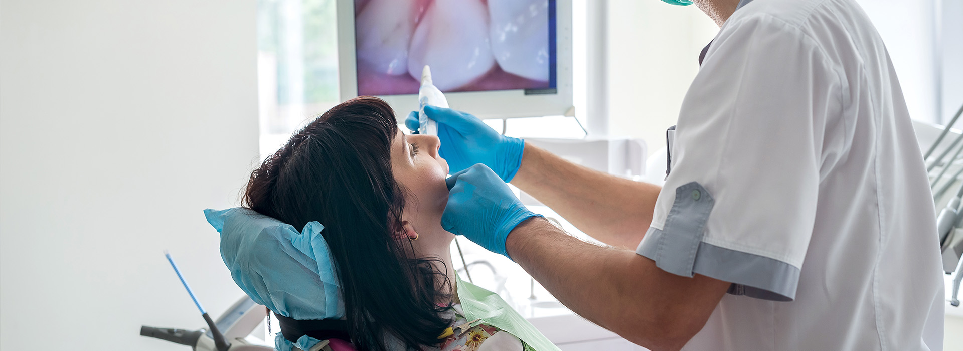
Dentist in blue gloves uses an intraoral camera on a reclined patient while a monitor shows an enlarged view of her teeth

An intraoral camera is a miniaturized camera that can take high-resolution images inside of the mouth and display them for viewing in real time on a computer screen. It is a pen-sized device that provides an up close, full-color and high-resolution view of the teeth and surrounding soft tissues.

An intraoral camera is a wonderful communication tool that allows the dentist to directly show a patient a close up view of every tooth and the nearby soft tissues. With this technology any areas of concern can be instantly displayed and discussed.

The intraoral camera can also capture individual images that can be saved as part of a patient’s permanent record. These saved images can be reproduced when needed for use by other dental specialists, a dental laboratory, insurance companies, and others.

Frequently Asked Questions

What is an intraoral camera and how does it work?

+

An intraoral camera is a compact, handheld imaging device that captures high-resolution stills and live video of teeth, gums, and other oral structures. It typically contains a small sensor, focused light source, and optics that allow clinicians to photograph areas that are difficult to see with the naked eye. The device connects to a computer or monitor so images appear in real time during an exam.

Modern intraoral cameras often include features such as autofocus, freeze-frame capture, and simple annotation tools to highlight areas of interest. Because the camera produces full-color images, it is especially useful for documenting visible surface conditions, guiding conversations about oral health, and creating visual records. It complements other diagnostic tools rather than replacing radiographs or 3D scans.

How does an intraoral camera improve diagnosis and treatment planning?

+

High-resolution intraoral images reveal subtle enamel changes, small fractures, staining, and soft-tissue abnormalities that might be missed during a quick visual exam. Enlarged and illuminated views help clinicians inspect restoration margins and hard-to-see molars with greater confidence, which supports more accurate documentation and prioritization of care. Captured images become part of a clinical record that clinicians can review when developing treatment plans.

Images can be compared over time to monitor progression or healing and help clinicians choose conservative, evidence-based interventions when appropriate. They also inform decisions about when additional imaging, such as radiographs or CBCT, is necessary to assess deeper structures. At Macri Dental, intraoral photography is incorporated into the diagnostic workflow to support clear, consistent treatment planning.

Will I feel any discomfort when an intraoral camera is used?

+

Use of an intraoral camera is generally noninvasive and causes minimal to no discomfort for most patients. The device is roughly the size of a pen and is designed to be maneuverable so clinicians can access different areas of the mouth with gentle movements. Patients who have a strong gag reflex or limited mouth opening should mention that to the clinician so adjustments can be made to improve comfort.

Clinicians commonly use single-use sheaths or barriers when operating the camera, which adds a thin layer between the device and oral tissues and does not interfere with imaging quality. The procedure is quick and often completed in seconds for each view, making it suitable for routine exams as well as specific diagnostic checks. If any discomfort is felt, the dental team can pause and modify technique to keep the exam comfortable.

How are intraoral images used to educate patients about their oral health?

+

Intraoral images transform abstract explanations into concrete visuals by showing patients close-up views of plaque, enamel wear, inflamed tissue, or restoration margins. Seeing a real-time image of a concern helps patients understand the nature and urgency of an issue and makes it easier to discuss recommended home care or professional treatment. Clinicians can annotate images to point out specific areas and explain the relationship between observed problems and daily habits.

Because images can be archived and compared, clinicians can illustrate progress or response to treatment during follow-up visits, which reinforces patient learning. Clear visual evidence often improves patient engagement and helps align expectations before treatment begins. This visual approach supports more informed decision-making and encourages adherence to preventive recommendations.

How does an intraoral camera integrate with other digital dental technologies?

+

Intraoral cameras integrate smoothly into modern digital workflows by linking captured photos with digital radiographs, intraoral scans, and treatment-planning software. Combined records create a more comprehensive, patient-centered view that clinicians can use to coordinate care and plan restorations with greater precision. Interoperability allows images to be displayed alongside other diagnostic data during consultations, which streamlines clinical presentations and decision-making.

When intraoral photography is used in tandem with radiographs and 3D scans, clinicians gain both surface detail and internal structure information needed for complex cases. This multi-modal approach supports predictable restorative work, implant planning, and monitoring of periodontal conditions. The combined datasets improve communication within the care team and enhance the overall quality of clinical records.

What are the limitations of an intraoral camera compared with X-rays or CBCT?

+

An intraoral camera excels at documenting surface details but cannot visualize bone, tooth roots, or internal anatomy the way radiographs or CBCT can. It is primarily a tool for recording visible conditions—such as enamel defects, soft-tissue changes, and restoration margins—rather than diagnosing subsurface pathology. For assessing cavities between teeth, bone loss, or root structure, clinicians rely on radiographic or three-dimensional imaging modalities.

Because of these limitations, intraoral imaging is most effective when used as part of a broader diagnostic protocol that includes radiographs or CBCT when indicated. Clinicians use photos to guide initial assessments and communication while ordering additional imaging when internal evaluation or 3D context is required. This complementary approach yields a more complete and accurate diagnosis than any single tool alone.

How are images from an intraoral camera stored and protected in my dental record?

+

Images captured during an exam become part of the patient’s clinical record and are stored in secure, practice-managed imaging systems alongside radiographs and notes. Access to these records is restricted to authorized members of the care team and used for clinical decision-making, continuity of care, and documentation of treatment outcomes. Proper recordkeeping ensures that images remain available for future comparison, referral, or laboratory communication when needed.

When images need to be shared with specialists or a dental laboratory, clinicians follow privacy and consent practices to protect patient information. Secure transfer methods and limited disclosure minimize risk while enabling collaborative care. Images retained in the chart also support quality control and efficient case presentation during interdisciplinary treatment planning at Macri Dental.

What infection control measures are used with intraoral cameras?

+

Infection control for intraoral cameras typically includes the use of single-use plastic sheaths or barriers and cleaning protocols recommended by the manufacturer and regulatory guidelines. Barriers prevent direct contact between the device and mucous membranes, and the camera is wiped or disinfected between patients according to the practice’s sterilization policies. Routine maintenance and adherence to established procedures keep the equipment hygienic and functioning properly.

Staff are trained to handle, sheath, and clean intraoral cameras consistently to protect patients and maintain image quality. These noninvasive devices are safe for routine use across most patient populations when infection-control practices are followed. Clear protocols also reduce downtime and ensure reliable performance for diagnostic imaging during appointments.

Who benefits most from intraoral imaging during dental care?

+

Many patient groups gain value from intraoral imaging, including those undergoing restorative work, patients with periodontal concerns, individuals considering cosmetic treatment, and parents of young children. The visual detail helps clinicians plan precise restorations, assess the condition of existing work, and communicate specific hygiene needs to patients and caregivers. Patients who prefer to see visual evidence of an issue often find it easier to participate in care decisions when images are used.

Intraoral cameras are also helpful for monitoring high-risk areas and tracking changes over time, which benefits patients with active disease or those receiving long-term maintenance. The technology supports tailored treatment strategies and enhances preventive counseling for a wide range of ages and clinical conditions. By improving transparency and involvement, intraoral imaging contributes to better-informed patients and more targeted clinical care.

How do intraoral images help when collaborating with specialists or dental laboratories?

+

High-quality intraoral photographs reduce ambiguity when communicating with specialists or dental laboratories by showing exact surface details, shade relationships, and restoration margins. Laboratories can use close-up views to match contours and shades more accurately, while specialists reviewing a referral gain a clearer picture of the clinical presentation before a consultation. Sharing images expedites case evaluation and helps ensure that all parties understand the same visual information.

When combined with radiographs and digital scans, photographic records provide a fuller context for treatment planning and lab fabrication. Clear visuals support timely, precise collaboration and help avoid unnecessary delays or repeat appointments. Well-documented intraoral images therefore improve the efficiency and predictability of interdisciplinary dental care.

Contact Us

Exceptional Dentistry, Centered Around You

Discover a dental experience built around your comfort, needs, and goals. Our team is here to answer your questions, help you explore your treatment options, and make scheduling your visit simple and convenient. Contact our office today and take the next step toward personalized, high-quality dental care for you and your family.

Office Hours

Monday
9:00 am - 5:00 pm
Tuesday
7:00 am - 4:00 pm
Wednesday
7:00 am - 3:00 pm
Thursday
7:00 am - 2:00 pm