Smiling woman with a blonde bob and tan cardigan showing healthy teeth after periodontal treatment

Digital Implant Planning

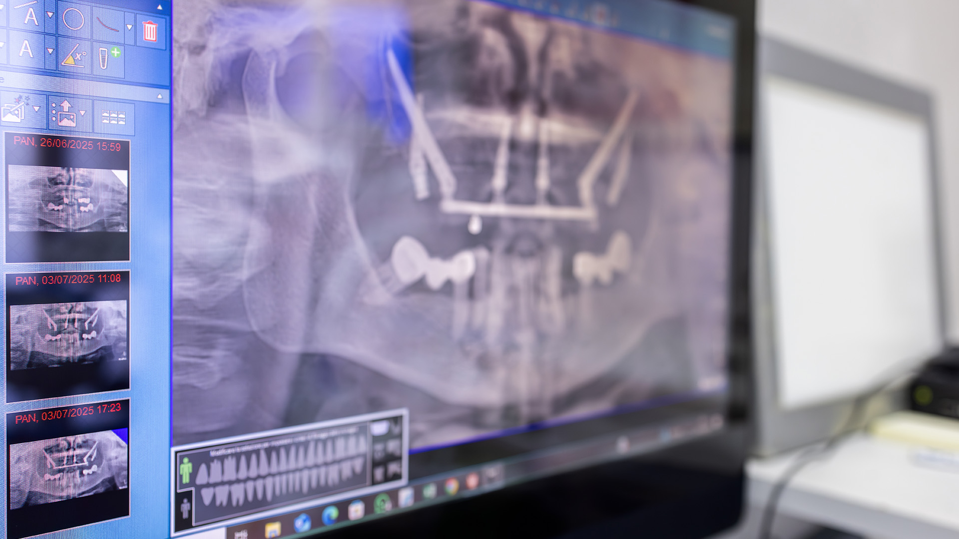
Computer screen showing a panoramic dental x-ray of a jaw with implant planning guides and thumbnail scans beside it

Why Digital Implant Planning Matters

Digital implant planning transforms what used to be a largely guess-driven process into a methodical, patient-specific workflow. Rather than relying solely on experience and two-dimensional X-rays, modern planning uses three-dimensional imaging and software to visualize the jaw, nerves, and surrounding anatomy before any incision is made. This shift from intuition to data-driven decisions helps the clinical team choose the exact implant position, angulation, and size that best fits each patient’s unique anatomy.

For patients, that means fewer surprises and a clearer understanding of the proposed treatment. Digital planning allows clinicians to anticipate potential challenges—such as limited bone volume or proximity to vital structures—and to design predictable solutions in advance. The result is a more streamlined surgical experience and a higher likelihood that the implant will integrate successfully and support a functional, natural-looking restoration.

Clinically, digital planning supports better communication across the treatment team. Surgeons, restorative dentists, and lab technicians can all review the same virtual model, discuss options, and agree on the ideal outcome. When everyone is working from the same plan, transitions between phases—planning, placement, and restoration—become smoother and more efficient.

State-of-the-Art Imaging and 3D Analysis

At the heart of digital implant planning is high-resolution, three-dimensional imaging. Cone beam computed tomography (CBCT) captures the jawbone in fine detail, showing bone height, width, and internal structures that traditional X-rays cannot. Advanced software then reconstructs these images into interactive models, enabling the clinician to examine cross-sections, measure critical distances, and assess bone quality with precision.

Beyond static images, software tools can simulate the position of virtual implants within the jaw. These simulations make it possible to trial multiple treatment scenarios—different implant sizes, angulations, and restorative positions—without any risk to the patient. The clinician can evaluate which option provides the best balance of long-term support, esthetics, and simplicity of restoration.

This imaging-driven approach also improves patient education. Visualizing the problem and the planned solution helps patients understand why a particular approach is recommended, what the surgical steps will look like, and how the final restoration will align with their bite and smile. Clear visuals reduce uncertainty and help patients make informed decisions about their care.

Precision from Virtual Surgery to the Chair

Digital implant planning is more than preoperative analysis; it enables physical tools that translate the virtual plan into accurate surgical outcomes. Using the digital plan, clinicians can design and fabricate surgical guides—custom templates that fit over the teeth or gums and guide the implant drills to the exact planned position and depth. These guides significantly reduce the variability that can occur during freehand placement.

Whether implants are placed immediately after extraction or in healed sites, the ability to carry a virtual plan into the operatory improves reproducibility and reduces intraoperative decision-making. For complex cases, guided surgery can decrease chair time and help maintain consistent implant alignment, which is critical for long-term function and for creating restorations that fit precisely on the first try.

Furthermore, the integration between digital planning and restorative workflows supports efficient prosthetic outcomes. When the planned implant position considers the final crown or bridge from the outset, the restorative phase is more predictable, and fewer adjustments are needed. That coordination preserves healthy tissue, improves esthetic outcomes, and often streamlines the number of appointments required to complete treatment.

How Digital Planning Improves Safety and Predictability

Safety is a central benefit of digital implant planning. Three-dimensional visualization helps clinicians identify and avoid critical anatomical structures—such as the inferior alveolar nerve in the lower jaw or the sinus cavity in the upper jaw—before the first incision. By mapping these structures relative to the planned implant, the surgical approach can be adapted to minimize risk and preserve function.

Predictability comes from reducing uncertainty at every stage. Preoperative planning lets surgeons select implants with appropriate length, diameter, and thread design based on measured bone dimensions. It also allows for contingency planning if the available bone is marginal, so augmentation or alternative strategies can be arranged in advance rather than decided under time pressure during surgery.

In addition to reducing intraoperative surprises, digital records create a reliable archive of the treatment rationale. Virtual plans, images, and measurements can be reviewed in the future if maintenance, repair, or replacement becomes necessary. That continuity of information helps maintain long-term outcomes and supports proactive follow-up care.

What to Expect During Your Digital Implant Consultation

A typical digital implant consultation begins with a thorough clinical exam and a discussion of your goals. If implants are an appropriate option, a CBCT scan is usually taken to capture the three-dimensional anatomy. The clinician then uses planning software to analyze the images and build a virtual plan tailored to your mouth, bite, and desired esthetic result.

During the planning stage, you can expect a clear explanation of the proposed implant position and the steps involved in surgery and restoration. The team will walk you through the logic behind implant selection and placement, including how the plan addresses concerns like bone support and nerve safety. This conversation helps align expectations and ensures the plan suits both functional and cosmetic objectives.

Finally, the discussion will cover the practical timeline and what to expect on the day of surgery—without getting into technical pricing or consultative offers. If a surgical guide or additional procedures are recommended, the clinician will explain how those elements fit into the treatment sequence. Throughout, the focus remains on creating a care plan that is precise, safe, and tailored to your long-term oral health.

Next Steps and How We Help

Digital implant planning is a collaborative, technology-driven process that brings clarity to complex treatment decisions. By combining advanced imaging, specialized software, and clinical expertise, it helps clinicians design treatments that are safer, more predictable, and better aligned with each patient’s goals. When planning is thorough, implant surgery and restoration proceed more efficiently and with greater confidence.

If you’re considering implants and want to learn how digital planning could benefit your case, our team is available to discuss the technology and the clinical considerations specific to your situation. Dr. Macri and the clinical staff bring extensive experience in digital workflows and implant placement, and they prioritize clear communication and thoughtful planning at every step.

Please contact us to request more information or to schedule a consultation. We would be glad to explain how digital implant planning can improve the predictability and outcome of your treatment.


Frequently Asked Questions

What is digital implant planning and why does it matter?

+

Digital implant planning is a data-driven workflow that uses three-dimensional imaging and specialized software to map a patient’s jaw, nerves and surrounding anatomy before any incision is made. By visualizing bone volume, tooth position and occlusion in a virtual model, clinicians can select the exact implant position, angulation and size that best match each patient’s anatomy. This shift from two-dimensional guesswork to precise planning leads to fewer intraoperative surprises and more predictable long-term outcomes.

The process also improves patient communication by offering clear visual explanations of the proposed treatment and expected results. Patients gain a better understanding of why a particular approach is recommended and what the surgical and restorative steps will involve. That transparency helps align expectations and supports informed consent.

How does CBCT imaging improve implant planning?

+

Cone beam computed tomography (CBCT) captures high-resolution, three-dimensional images of the jaws that reveal bone height, width and internal structures that conventional X-rays cannot show. These volumetric images allow clinicians to measure critical distances to nerves and sinus cavities and to evaluate bone quality at potential implant sites. With this level of detail, clinicians can make more confident selections of implant length, diameter and trajectory.

CBCT data can be imported into planning software to create interactive models and cross-sectional views for precise analysis. The models support simulations of multiple treatment scenarios so clinicians can compare restorative and surgical options without risk to the patient. This reduces uncertainty and helps the team prepare for potential augmentation or alternative strategies before entering the operatory.

What is a surgical guide and how does it improve guided implant surgery?

+

A surgical guide is a custom-fabricated template derived from the digital implant plan that fits over the teeth or gums and directs drill position, angulation and depth during surgery. Because the guide is manufactured to match the virtual plan exactly, it translates the planned implant coordinates into the clinical field with high fidelity. This reduces the variability common with freehand placement and helps achieve the intended implant orientation and depth.

Surgical guides are especially valuable in complex or esthetic cases where precise implant alignment is critical for prosthetic success. They can decrease chair time, limit intraoperative decision-making and increase reproducibility across cases. When the surgeon and restorative team coordinate the guide design, the result is a smoother transition from placement to restoration.

Who is a good candidate for digital implant planning?

+

Most patients being considered for dental implants can benefit from digital implant planning, particularly those with limited bone volume, complex anatomy or proximity to vital structures such as the inferior alveolar nerve or the sinus cavity. Patients with single-tooth replacements, full-arch reconstructions or prior grafting requirements also gain value from the increased precision and forethought the process provides. The technology is useful across a wide range of clinical scenarios because it supports individualized treatment decisions.

During an initial evaluation, the clinician will review medical and dental history, perform a clinical exam and, when appropriate, obtain a CBCT scan to assess candidacy. If the scan and examination indicate implants are feasible, the team will develop a digital treatment plan that addresses bone support, prosthetic needs and any adjunctive procedures. The result is a tailored approach that considers both surgical and restorative objectives.

How does digital planning improve safety and reduce surgical surprises?

+

Digital planning enhances safety by allowing clinicians to identify and map critical anatomical landmarks before surgery, reducing the risk of damage to nerves, blood vessels or sinus membranes. Measurements taken from three-dimensional images guide implant selection and depth control, and potential conflicts with vital structures can be detected and managed during planning rather than in the operatory. This preoperative foresight minimizes unplanned intraoperative changes and supports a more controlled surgical environment.

In addition to anatomical assessment, digital planning lets clinicians anticipate whether bone grafting, sinus lift or other adjunctive procedures will be necessary. Contingency plans can be prepared in advance so that appropriate materials, instruments and scheduling are in place. The cumulative effect is a lower likelihood of unexpected complications and a smoother treatment course for the patient.

How does digital planning affect restorative outcomes and prosthetic fit?

+

When implant position is planned with the final restoration in mind, the restorative phase becomes more predictable and efficient. Digital workflows allow the team to simulate restorative emergence profiles, crown height and occlusion during the planning phase so the implant is placed to support an optimal prosthetic outcome. This forward planning reduces the need for major adjustments to crowns or abutments and helps preserve healthy soft tissue contours.

Integration with digital lab workflows and 3D printing further streamlines prosthetic fabrication and verification. Labs and technicians can work from the same virtual plan used for surgery, which improves communication and reduces remakes. The result is restorations that fit more accurately on the first try and better long-term esthetics and function.

What should I expect during a digital implant consultation?

+

A typical digital implant consultation begins with a comprehensive review of your medical and dental history, a clinical exam and discussion of your goals for function and esthetics. If implants are appropriate, the clinician will usually take a CBCT scan and digital impressions to capture the three-dimensional anatomy and occlusion. These records are then evaluated with planning software to create a customized virtual plan tailored to your mouth.

During the consultation the clinician will explain the chosen implant position and the rationale behind implant type, angulation and restorative considerations. You will also be informed about the sequence of treatment steps, whether a surgical guide is recommended and what to expect during surgery and restoration. The focus is on clear communication so you understand the plan and the anticipated clinical timeline.

How does the care team coordinate using digital workflows?

+

Digital implant planning promotes multidisciplinary collaboration by providing a shared virtual model that surgeons, restorative dentists and laboratory technicians can review together. The unified dataset enables precise discussion of implant placement, prosthetic contours and sequencing, which reduces miscommunication and aligns goals across providers. When everyone works from the same plan, transitions between planning, placement and restoration are more efficient and predictable.

At a practice like Macri Dental, clinicians with implant training and experienced support staff integrate digital tools into routine workflows to ensure consistent outcomes. Files from the planning software can be exported for guide fabrication or to a dental lab, streamlining the production of surgical templates and prosthetic components. This coordinated approach helps maintain quality control from the virtual plan through final restoration.

How should I prepare for implant surgery that uses digital planning?

+

Preparation for guided implant surgery typically involves standard preoperative steps such as reviewing medical history, discontinuing or adjusting certain medications under medical guidance and arranging transportation if sedation will be used. You may be asked to follow fasting guidelines if sedation is planned and to coordinate any required medical clearances. The digital plan itself reduces unexpected intraoperative decisions, which can shorten operative time and simplify preparation.

Your clinician will provide specific instructions about day-of-surgery expectations, postoperative care and any temporary restorations that may be placed. Because the surgical guide and implant components are prepared in advance, the team can focus on efficient, precise execution on the day of surgery. Clear preoperative communication helps ensure a smoother appointment and recovery.

What kind of follow-up and maintenance are recommended after implant placement?

+

After implant placement, initial follow-up visits focus on monitoring healing, assessing soft tissue response and confirming integration of the implant with the bone. The restorative phase will proceed once the clinician determines the implant is stable, and periodic follow-up appointments will be scheduled to check occlusion, tissue health and prosthetic function. Good oral hygiene and routine dental visits are essential to maintain implant health over the long term.

Long-term maintenance typically includes professional cleanings, radiographic evaluations as indicated, and patient education on home care techniques for implants and surrounding tissue. Digital records and archived plans serve as a useful reference for future maintenance, repair or replacement needs. Consistent monitoring and prompt attention to any changes help preserve function and esthetics for years to come.

Contact Us

Exceptional Dentistry, Centered Around You

Discover a dental experience built around your comfort, needs, and goals. Our team is here to answer your questions, help you explore your treatment options, and make scheduling your visit simple and convenient. Contact our office today and take the next step toward personalized, high-quality dental care for you and your family.

Office Hours

Monday
9:00 am - 5:00 pm
Tuesday
7:00 am - 4:00 pm
Wednesday
7:00 am - 3:00 pm
Thursday
7:00 am - 2:00 pm